
Hip wear is the most common form of osteoarthritis. All information on causes, typical symptoms, treatment options and prevention.
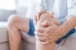
- Degenerative changes in the hip joint cause pain, the main symptom of hip joint osteoarthritis, also called coxarthrosis.
- © iStock.com/itsmejust
Coxarthrosis (alternative terms: hip arthrosis or hip joint arthrosis) is the medical term for the change in the joint on the hip due to wear and aging (degeneration). The joint wear leads to degradation and ultimately destruction of the cartilage in the hip joint.
In addition to knee arthrosis, coxarthrosis is one of the most common types of arthrosis. An estimated five percent of adults in Germany suffer from painful wear and tear of the hip joint, and the frequency increases with age.
Relieve pain in osteoarthritis

Hip arthrosis causes considerable costs in the health care system. The severely restricted mobility often requires an operation in which the patient is given an artificial hip joint. According to figures from the OECD, an "artificial hip" was used around 230,000 times in Germany in 2013. With the right measures, surgery can be avoided for a long time.
Causes and risk factors for coxarthrosis
The causes of hip arthrosis include congenital or acquired joint misalignments, nutritional disorders of the articular cartilage, incorrect stress, joint injuries or broken bones as well as inherited predispositions.
An overview of the possible causes of coxarthrosis:
congenital malformations of the hip joint like hip dysplasia
Childhood diseases like the youthful hip head solution or the Perthes disease, In the latter, the joint head is not sufficiently supplied with blood, dies and deforms.
Hip deformities (for example rachitic bow legs or X-legs) or long-term heavy load or incorrect load, which leads to an uneven pressure distribution within the joint
joint injuries, Broken bones, especially in the area of the femoral neck
Disorders in joint metabolism (non-bacterial inflammation, rheumatism, bacterial inflammation): In these diseases, inflammatory processes or deposits of metabolic products destroy the inner skin of the joint or the cartilage.
overweight accelerates changes in the weight-bearing joints as the body weight increases the pressure on the cartilage.
heredity with changes in hip shape or cartilage quality
Congenital malformation: hip dysplasia
Mostly due to an inherited disposition, but also partly due to a special position of the embryo in the uterus, the so-called acetabular roof, which closely surrounds the femoral head, can be incomplete. This allows the joint head to slide upwards and, in extreme cases, come out completely from the socket (hip dislocation). The acetabular roof is excessively stressed and is particularly sensitive to the development of hip arthrosis. Hip dysplasia, which is undetected in childhood and not treated, is referred to as pre-arthrosis (precursor to arthrosis).
Perthes disease (Perthes disease)
Disorders of cartilage and bone growth can lead to Perthes disease in childhood and adolescence. Here the femoral head dies. If it is loaded, it deforms. The organism can rebuild the joint head to a certain extent. If, however, he is put under too much strain under this new construction, the joint head will no longer gain the round shape necessary for smooth joint play; because only a smooth hip joint is a good joint.
Joint deformities
Ideal axes exist for each joint. If the joint is in the ideal axis, the forces applied to the joint are optimally distributed. If the joint position is disturbed, the load increases at certain points and cartilage abrasion occurs. Incorrect loads in distant joints (for example, the big toe joint, knees) also affect the hip. Pain in the foot or hip can protect one leg, and the other hip is then put under more strain. The risk of coxarthrosis increases.
Joint injuries, broken bones
Every change in shape reduces joint function. In the event of a fracture of the femoral neck, in which the femoral head is separated from the rest of the femur, the mobility of the joints is severely restricted during the healing process and the risk of malnutrition of the cartilage with subsequent osteoarthritis is high.
Non-bacterial joint inflammation: osteoarthritis due to rheumatism
Osteoarthritis and rheumatism are two different clinical pictures, but are often confused. However, osteoarthritis can result from rheumatism.
Osteoarthritis initially means only joint wear due to increased or incorrect stress. The activated arthrosis is an inflammation, whereby the inflammatory stimulus is not a pathogen and not a defense reaction, but a foreign body (e.g. rubbed-off cartilage particles).
Rheumatism (primarily chronic polyarthritis) is a disease in which the immune system is directed against the body's own cells because it believes that these cells are foreign to the body due to incorrect information. This type of immune dysfunction is known as autoaggressive disease. The cause here is not the joint itself, but a misguided defense reaction. In rheumatism, the cells of the inner skin of the joint are the target of the wrong immune response.
Ten tips for joint pain

Due to the immune process, which ultimately also manifests itself as inflammation, the inner skin of the joint thickens. This also disrupts the joint function so that rheumatism can result in arthrosis of the hip joint. Two joint-destroying processes come together here, so that the original disease – rheumatism – must be treated intensively. It is typical of rheumatism that this disease affects several joints. A change that is limited to the hip joint is usually not rheumatism.
Bacterial inflammation
Bacteria carried over to the blood can settle and spread in the synovial fluid (synovia). There they lead to bacterial joint inflammation. The metabolism is greatly increased in the case of inflammation (recognizable by the reddening and overheating). There are an increasing number of metabolic end products that can only be removed with a delay from the joint gap that slowly participates in the general metabolism. The mainly acidic waste products can lead to sustainable cartilage damage. Bacterial colonization from the blood is less common today.
Bacterial joint inflammation can also occur after a joint puncture or joint operation. Most of all one Longer cortisone treatment of the joints and diabetes (diabetes mellitus) reduce the immune system against pathogens and thus increase the risk of cartilage damage and joint wear.
You can find out more about the causes of the various forms of osteoarthritis here.
Symptoms: How the hip osteoarthritis becomes noticeable
The symptoms of hip joint osteoarthritis are so typical that an x-ray is usually necessary to ensure the diagnosis.
Significant signs of illness are hip pain, restricted movement and muscle tension in the area of the affected joint. With activated arthrosis also come signs of inflammation such as swelling, redness and overheating. They are also a typical sign of advanced osteoarthritis crepitus,
Pain course in coxarthrosis
Almost all patients (95 percent) with hip osteoarthritis state that the pain initially occurs when the movement begins start-up pain occurs. After a long period of rest as well as in the morning a feeling of rigidity join in. The start-up pain initially disappears after a short time and only occurs when the load is longer fatigue pain again. When the osteoarthritis progresses, the pain finally persists.
For hip arthrosis this is typically Get up very painful from a deep armchair; the hip pain often pulls from the groin to the thigh.
-
Signs of wear on the joints that are accompanied by pain are called osteoarthritis. Are you at risk? Test yourself!
for self-test
Muscle tension by protecting the painful joint
The restriction of movement is initially due to pain. The patient tries to close the joint save and avoid particularly painful movements. This leads to reflexive, i.e. involuntary muscle tension, which in turn causes further pain. Muscle tension that lasts longer (more than two weeks) ends in shortening (contractures) of the muscles and further restrict the possible range of motion.
The doctor also assesses passive mobility in the joint. With hip arthrosis, the thigh Bend and stretch to a limited extent, only spread under pain and turn poorly or not at all.
Joint effusion can initially relieve pain
When arthrosis is activated, it swells joint capsule which leads to a feeling of tension in the groin. With a chronically creeping degeneration the joint is through osteophytes also thicker, but this is hardly noticeable under the muscles.
Due to the irritation of the Synovium Rubbed cartilage particles lead to inflammatory changes in the area of the affected joint. The joint capsule is swollen and it can also be a so-called Joint effusion occur. This increased accumulation of fluid in the joint cavity often leads to a temporary one pain relief, because the articular surfaces slide better together.
A decrease in pain is not necessarily a sign of one degeneration the coxarthrosis, but rather can be an indication of a joint effusion.
Rubbing noises due to abrasion from cartilage particles
Abrasion from cartilage particles and the roughened cartilage surface sometimes lead to another symptom of hip arthrosis: rubbing noises can be heard or felt over the joint crunch be felt when touching the joint.
You can find out more about the general symptoms of osteoarthritis here.
This is how the doctor diagnoses osteoarthritis of the hip
After collecting the medical history, the physical examination and laboratory examinations, the doctor has a number of equipment options to diagnose coxarthrosis. They include X-ray technology, ultrasound examinations (sonography) and scintigraphy.
At the beginning of the examinations in the case of suspected arthrosis of the hip joint, the medical history is recorded by the doctor. He asks about the type and duration of the complaints, accompanying diseases and joint diseases in the patient's family.
This is followed by a physical examination, in which attention is paid to external changes in the shape of the hip joint and signs of inflammation such as redness, swelling and overheating. The mobility is examined and the doctor looks at the gait pattern of the patient. He can also recognize malpositions of the joint axes, which can be the cause of the joint disease.
X-ray
The doctor can already see the extent of the damage to the cartilage in the hip, the malposition of the hip joint and the alteration of the cartilage structure on the X-ray image. The cartilage is translucent so that it appears as a gap between the joint parts. A worn, thin cartilage can be seen as a thinner gap compared to the normal one.
blood tests
In order to distinguish a pure wear of the hip joint from an inflammation, which has to be treated differently, the blood can be examined for inflammation values. Accelerated blood cell lowering reactions, an increased level of C-reactive protein (CRP value, which increases rapidly with inflammation) and a typically changed electrophoresis indicate inflammation in the body.
Other laboratory values that can be examined are:
A bacterial culture is also created to rule out bacterial streptococcal infections.
Ultrasound examinations (sonography)
The soft tissues (muscles, tendons, ligaments) and fluid accumulations in the joints are permeable to X-rays, so they cannot be shown in the X-ray image. On the other hand, they are clearly visible through ultrasound. However, the resolution of this image-giving process is not too high on the joint.
scintigraphy
The question "inflammatory or degenerative?" the doctor can answer using scintigraphy. In this investigation, weakly radioactive substances are injected into the vein. The substances accumulate particularly in the bones or in the soft parts. Inflammatory tissue or tissue that has changed due to malignant growths enriches the substance more than healthy tissue, so that these areas are clearly recognizable.
Computed tomography and magnetic resonance imaging
In some cases, computed tomography (CT) or magnetic resonance imaging (MRI) is useful before surgery because both procedures can provide very detailed slices. The MRI can be used to visualize ligaments, inner skin of the joint and cartilage.
You can get an overview of the general diagnosis options for osteoarthritis here.
From physiotherapy to hip prosthesis: treatment of coxarthrosis
Depending on the clinical picture, various treatment options can be considered for hip arthrosis. If the coxarthrosis is at rest, an attempt is made to maintain mobility with as little discomfort as possible. Only when the quality of life is significantly impaired by the pain and the restricted movement is an operation necessary.
The individual stages of treating coxarthrosis are:
joint-friendly lifestyle changes, These consist primarily in learning movement sequences that are gentle on the joints
physical treatment (Heat / cold, protection, physiotherapy, massages, electrotherapy, ultrasound therapy). The patient can use some of these methods at home as part of self-help, others are carried out by physiotherapists
relieves pain and inflammation drugs
joint-preserving operations or replacement of the hip joint
Protection and relief with activated hip arthrosis
At a activated coxarthrosis with signs of inflammation in the joint (overheating or reddening) you should perceive the pain as a warning signal and protect the joint as best as possible. Sitting with a half-raised upper body and high legs is gentle on the hips; slightly bent knees (pillow or bolster underlay) are favorable. If a few steps cannot be avoided, a stick or a forearm crutch are useful. Relief is also possible through soft shoe soles (buffer heels); because the bumps on occurrence are transmitted to the hip joint.
If the immobilization is extended too long, there is a great risk that the joint will stiffen. Please be sure to seek advice from your doctor when you can carefully and gradually load the joint again.
Cold soothes active inflammation
Cold dampens all metabolic processes, including inflammation processes with pain. at activated hip arthrosis with overheating relieves cold the complaints are effective. Cold envelopes can be made with (ice) water, cold packs or alcohol. However, these should not be placed directly on the skin (place a damp cloth underneath). The cold edition is changed as soon as it has warmed up. The application of cold should be pleasant and should not last longer than 20 minutes.
Warmth relaxes the muscles
Most patients with resting hip arthrosis find warmth to be very pleasant. The heat relaxes the muscles, the movements run more fluently and smoothly, which benefits the joints directly. Warm pads and warmth in the form of heating pads, infrared lamps (be careful: protect your eyes!), Heated gel packs, mud packs and thermal baths provide relief. Even a warm bath at home relaxes. This effect can be promoted by bath additives, especially of a vegetable type (lavender, lemon balm). If you find rheumatism old-fashioned, you can look for ski underwear in sports shops.
Pain can also be due to inflammation that is worsened by heat. Therefore, do not apply heat directly on the joint, but over the neighboring muscles!
Physiotherapy to strengthen the muscles
Physiotherapy is not a substitute for active movement, but should specifically strengthen the muscle coat around the joints in order to protect and relieve the hip. It is of fundamental importance in physiotherapy that you do the exercises at home regularly and over a long period of time. The few medically prescribed physiotherapy lessons are not the whole program. They should be understood as instructions so that you know what you can do at home to improve the symptoms of hip arthrosis very significantly.
Exercise: swimming or cycling regularly
According to the motto "move more, burden less", patients with coxarthrosis should regularly practice a sport such as swimming or cycling. The entire weight of the upper body does not rest on the hip joints, which can be moved freely – even in the water supported by buoyancy. The movement improves the supply of nutrients to the articular cartilage and the lubricity of the synovial fluid. However, movements under increased stress, such as mountaineering, tennis and skiing, should be avoided as far as possible.
Two exercises for hip pain caused by osteoarthritis
Liebscher & Bracht / YouTube
Medication
There are two large groups of drugs available for coxarthrosis: cartilage-protecting or building-up preparations (chondroprotectives) and pain and anti-inflammatory agents (anti-rheumatic drugs). The latter can also be divided into two larger groups: non-steroidal and steroidal. Steroidal means "derived from steroids". These funds are essentially medicines from the cortisone family. Non-steroidal are drugs that affect inflammatory metabolism: acetylsalicylic acid (ASA) and relatives.
The aim of treatment with chondroprotectives is to heal the joint changes, the aim of treatment with the anti-inflammatory drugs is to eliminate the symptoms.
For some herbal remedies, for example rose hip powder or ginger, as well as for the supply of the protein glucosamine, which is part of the cartilage and connective tissue, the effectiveness of pain relief and improved mobility in osteoarthritis has been proven in studies.
Surgery for hip arthrosis
There are two types of operations:
Change of the joint axis by changing the thigh bone or the acetabulum (preserving the joint: corrective osteotomy)
Replacement of the arthrotic hip joint with an artificial joint, consisting of an artificial joint socket and an artificial femoral head (total endoprosthesis), rarely with an exclusive head endoprosthesis (KEP)
Principle of corrective osteotomy
The joint-preserving corrective osteotomy is intended to prevent osteoarthritis of the hip joint by changing the joint conditions so that the stresses are distributed more evenly and arthrosis does not develop at all or avoid further deterioration.
In this operation, the joint itself is not touched, only the shape of the thigh neck is changed or the acetabulum is pivoted over the femoral head. The thigh bone or the acetabulum must be sawed apart and reassembled. Screws and plates fix the new position. They are removed again when the new bony connection has become sufficiently stable.
Principle of total hip replacement
During this procedure, part of your own, modified hip joint is removed and replaced by two implants – one for the socket and one for the femoral head with an anchoring in the femur. The material of the hip prosthesis is metal, plastic and / or ceramic.
Treatment of arthrosis – what helps with joint wear? All information is here!
This is how coxarthrosis can be prevented
Osteoarthritis of the hip can be prevented quite well. In the case of major malpositions, for example a congenital hip dysplasia that was discovered late, preventive measures maintain the quality of life for a long time and help to postpone surgery. This is also an advantage because no artificial hip joint will last forever, even if there is great progress in this area today.
The possible measures to prevent coxarthrosis include:
- the Avoiding incorrect loads through balancing sports with one-sided or only low occupational stress, or even shoe height compensation when there is a difference in leg length
- the Avoid overloading the joints, This includes the reduction of overweight, but also the throttling of overly ambitious sports programs.
- Regular exercise is beneficial for any form of osteoarthritis, This ensures good cartilage nutrition and a stable muscle coat that relieves the joints. Swimming and cycling are ideal because the movement is possible without high stress on the joints.
- Varied nutrition affects the composition of the cartilage-nourishing synovial fluid.
- In the event of accidental injuries to the joint, care should be taken to ensure that cartilage, bone and ligament portions are restored as completely as possible. Even permanent bone levels of just a few millimeters can lead to osteoarthritis years or decades later.
- In the event of malposition, the change osteotomy is performed operative prevention the hip arthrosis. This procedure changes the axis of the joint by changing the angle between the femoral shaft and neck. As a result, the joint is set back in its correct axis, so that the incorrect loading is ended. A notable arthrosis can be avoided in this way.