Retinography is a process which allows the fundus of the eye to be photographed (retina, vitreous body, macula). This is a non-invasive examination, which allows the study of the different layers of the retina. It can be prescribed to diagnose eye pathologies or to monitor the evolution of other diseases having an impact on the eye (diabetes, hypertension). Find out what a retinogram is, in which cases to prescribe it and how it is carried out.
Retinography: what is it?
Retinography is an ophthalmology examination which allows your specialist to analyze the structures located at the back of the eye, behind the lens, in other words the vitreous body, the macula (part in the center of the retina) and the retina. This involves taking digital photos, through the pupil, of your retina and your optic nerve, in order to be able to analyze them in detail, specifies the Sorbonne/Saint-Michel ophthalmological center. Depending on the type of camera used for examining the fundus of the eye, we distinguish between classic retinography (centered on the retina and the macula) and wide-field retinography which includes the entire retina, informs the Paris 17 ophthalmological center. This examination takes place without injection of contrast product or direct contact with your eye. It is therefore a non-invasive examination.
What are the indications for a retinography?
The reasons which may lead your ophthalmologist to prescribe a fundus examination may be the screening of certain retinal or optic nerve pathologies, or the monitoring of diseases already diagnosed. Among the pathologies that retinography can highlight or whose evolution it can monitor, we note age-related macular degeneration (degradation of the macula which can lead to blindness), glaucoma (progressive damage to the optic nerve) , retinal detachment, vascular occlusion or diabetic retinopathy, for example. But this ophthalmology examination also makes it possible to monitor the possible repercussions on the eye of more general diseases such as diabetes or high blood pressure.
How does a fundus examination take place?
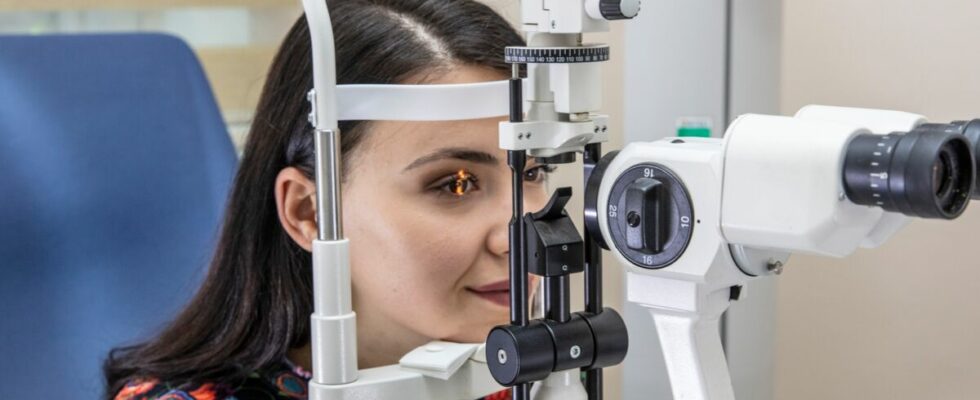
A fundus examination can be performed with or without dilation of the pupil beforehand. To dilate the pupil, the ophthalmologist applies mydriatic eye drops which take effect after 20 to 45 minutes of waiting. But this expansion is less and less necessary with new generation devices, even more precise and efficient than the previous ones. For the examination, your face is placed on a chin rest: this is to prevent you from moving and to keep your eye facing the lens. You are asked to focus on an image located in the center of the camera and a series of digital photos is then taken. The exam only lasts a few minutes.
Sources
- GlaucomaThe MSD Manual — Consumer Version, April 2023
- Macular degenerationNational Institute of Health and Medical Research (Inserm), June 14, 2017
- Retinography — Photography of the fundus of the eyeParis 17 Ophthalmological Center
- RetinographySorbonne Saint-Michel Ophthalmological Center, February 25, 2021
Read also :
⋙ Eye infection: the different symptoms to recognize and how to react
⋙ Spot in the eye: what are the causes of this vision disorder?
⋙ Eye cancer: symptoms, diagnosis and treatments for this rare disease