According to data from the French Association of Urology (Afu), Peyronie’s disease affects between 3% and 9% of men in France. To understand the mechanisms of this deformation of the penis during erection, urologists perform a penile MRI. This imaging examination is also useful before an intervention in the event of a penile fracture, to detect the origin of an erectile dysfunction or before the installation of a penile prosthesis.
Why does the urologist indicate a penile MRI?
The penis has a corpus spongiosum and two corpora cavernosa which give it elasticity and promote erection when they fill with blood. These bodies are surrounded by a white fibromuscular tunic which separates them, the albuginea. Like all other parts of the body, this complex mechanism can go wrong. This is when the urologist, a doctor specializing in the male genitals, kidneys and urinary tract, comes into play.
Penile MRI may be indicated in urology in the following cases:
- to investigate the cause of Peyronie’s disease, a pathology of the penis which is characterized by curvature during erection;
- if a patient complains of pain during erection or only during sexual intercourse (male dyspareunia);
- to locate the injury in the event of rupture of the corpora cavernosa (penile fracture);
- in case of penile cancer;
- in men with priapism (prolonged erection lasting several hours);
- before installing a penile implant.
How is a penile MRI performed?
On the day of the exam, you must give the medical imaging technicians the questionnaire that was given to you with the invitation. This questionnaire provides you with information on the procedure and its contraindications. A product used to treat erectile dysfunction, for example Edex or Caverject, will be injected into the corpora cavernosa. These products are contraindicated if you have sickle cell anemia (red blood cell abnormality of genetic origin) or multiple myeloma, or if you are allergic to prostaglandins (the injected product contains these molecules).
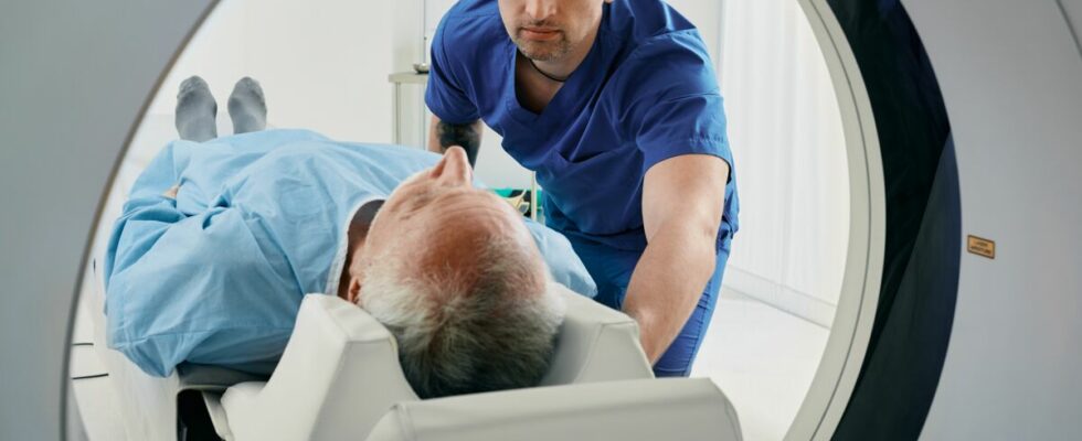
The intracavernous injection is performed approximately 10 minutes before the examination. It is not painful. Using a very fine needle, the radiologist injects a product which increases blood flow and causes an erection. A gadolinium-based contrast agent is also sometimes administered. With the erection, technicians will be able to obtain clearer and more precise images of the internal structure of the penis. You will lie on your back for 15 to 30 minutes. The images will be obtained using a small device, a surface antenna in contact with the penile region.
Can penile MRI cause side effects?
Complications are very rare. Some mild side effects may occur, for example, an allergic reaction and itching caused by the contrast agent (urticaria). There is also a risk of hematoma at the site of the injection, as with any injection. All metal objects carry a risk of burn or injury during the examination: pacemaker, piercing, artificial heart valve, hearing aid, dental ring, brooch, etc. If you are wearing an iron or metal object, you must indicate this during the examination. of making an appointment and the day of the exam. If you have a recent tattoo (less than 6 weeks), you must also report it.
What information does penile MRI reveal?
The information sought by the urologist varies depending on the disorders and the state of health of the person. If penile cancer is suspected, exploration is useful to confirm the presence of the tumor at an early stage, to localize its location and to prepare for surgery. For example, it allows us to know if the tumor has invaded the corpus spongiosum, the corpus cavernosum, the urethra or the prostate. In patients with Peyronie’s disease, it highlights plaques, nodules or calcifications inside the penis.
To treat priapism, penile MRI allows you to know whether it is a low-flow or high-flow form. This distinction is crucial, because urgent treatment must be implemented in cases of low-flow priapism. For penile fractures, no product is injected into the corpora cavernosa. The images reveal the location and extent of the fracture. If an injury to the urethra is visible, the urologist performs imaging of the urethra (urethrography).
Sources
- Peyronie’s disease, finally a treatment!press release from the French Association of Urology (Afu), September 13, 2016
- Treatment of Peyronie’s diseaseProgress in urology, journal of the French Association of Urology (Afu), 2009
- When and how to operate on rod fractures?Progress in urology, journal of the French Association of Urology (Afu), 2015
- MRI of the penisNational Library of Medicine, Dr Alex Kirkham, November 2012
- Magnetic resonance imaging (MRI) of the penisBordeaux University Hospital, March 2021
Read also :
⋙ MRI: indications and procedure of the examination
⋙ Scanner and MRI: what are the differences?
⋙ Exam: have an MRI without stress