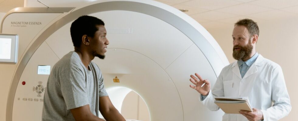
When we have an examination to do to study back pain, visceral discomfort, a problem that cannot be explained by blood tests or is not detected during consultation… We are sometimes referred to an x-ray, an ultrasound, or towards two other types of examinations, which are also field of medical imaging: scanner or MRI. How do they work? What are their differences ? Here are the answers to your questions.
Scanner and MRI: two medical imaging techniques
“An MRI (magnetic resonance imaging) is a radiology exam that uses a device emitting electromagnetic waves, using a large magnet. Subjected to these waves, the hydrogen atoms making up the body’s tissues begin to vibrate. They then emit signals, captured by a specific camera and transcribed into images on a computer screen. explains Health Insurance.
The scanner does not use the same technology as MRI: it “is made up of a ring equipped with an X-ray emitting tube and digital sensors which scan the area of the body to be studied by rotating. The device emits low-dose X-rays, directed towards the part of the body to be analyzed. It measures the absorption of X-rays, which differs depending on the nature of the tissues they pass through. The information is used to reconstruct an image of the body based on the ray absorption density. The scanner produces images in thin, serial anatomical sections (i.e. “thin slices”). These images are processed by a computer which allows a reconstruction in two or three dimensions (2D or 3D) of the areas of the body studied. complete Ameli.
MRI and CT: different duration and visualization
With the first, we do not undergo irradiation, with the second, we do. The scanner is therefore contraindicated in certain people, as in pregnant women. “In both cases the patient is installed in a tunnel but that of the MRI is much longer, which can cause discomfort in claustrophobic patients. The duration of the examination is also longer in MRI (around 30 minutes) than in CT (around 5-10 minutes),” highlights the radiology and imaging portal. The two exams share the common point of being painless.
These two types of medical imaging examinations therefore do not have the same approach, and are “complementary”, as explained on the Radiologie Paris Ouest website. They are not used to see exactly the same thing, nor in quite the same way. MRI makes it possible to obtain images of the inside of the body in two or three dimensions, it is useful for visualizing “soft tissues” such as the spinal cord, viscera, muscles or even the brain. It also allows you to see bones and joints. But it is the scanner which is more recommended for explore the bones, the ENT sphere, to visualize the deep organs of the abdomen and thorax. It makes it possible to detect various anomalies such as: “hemorrhage, tumor, cyst, infection, enlarged lymph node”, notes Health Insurance. It is used to “guide” cancer treatments, or when a puncture of deep organs is necessary.
Read also :
⋙ Ultrasound with a midwife: in which case?
⋙ Scanner with injection: how does the exam take place?
⋙ What is the outfit you should never wear to have an MRI?
