
An ECG is an indispensable diagnostic procedure for recording and analyzing the electrical activity of the heart. The non-invasive examination method provides valuable information about heart health. When is an ECG performed and how is it evaluated?
- © Getty Images/Westend61
Quick overview: ECG (electrocardiography)
Definition: EKG stands for electrocardiography. During the cardiological examination, the heart’s currents are measured and an ECG curve, the so-called electrocardiogram, is created.
When is an ECG necessary? An EKG is carried out if heart diseases such as cardiac arrhythmias or a heart attack are suspected.
What forms are there? In addition to the resting ECG, in which the measurement is taken while lying down and only takes a few seconds, there is also the stress ECG and the long-term ECG.
At a glance:
22 tips for a healthy heart

What is an ECG?
EKG is an abbreviation and stands for electrocardiography (alternatively: electrocardiography). The ECG is considered a basic cardiological examination with high informative value. This examination method is used to measure the electrical activity of the heart: changes provide important information about heart disease. The result of an electrocardiography is called an electrocardiogram.
For the ECG, electrodes are placed on the chest, arms and legs. They measure the electrical signal and record it. The voltage between two electrodes is compared; experts also speak of a derivation. The resulting electrocardiogram shows heart activity in the form of waveforms called “waves.”
Conduction of excitation in the heart: How does the heartbeat occur?
The heart pumps blood throughout the body, ensuring the vital supply of oxygen. To do this, it contracts rhythmically. These contractions are triggered by electrical impulses with a voltage of about a thousandth of a volt, which are delivered in a specific rhythm.
The electrical signal originates from the specialized cells of the sinoatrial node, which is located in the right atrium, and spreads through the atria and ventricles. The regular heartbeat that is generated in this way is therefore also known as sinus rhythm. When the electrical impulse has subsided, the heart muscles relax and then contract again with the next impulse.
This electrical activity of the heart follows a characteristic pattern that can be graphically displayed using electrocardiography in the form of an electrocardiogram. Deviations from this pattern indicate various illnesses.
When is an ECG performed?
Cardiac currents are measured when various heart diseases are suspected, including:
Even if there are conduction disorders in the heart, such as a bundle branch block or an AV block (interruption of impulse transmission in the heart), an ECG can reveal this.
Carrying out the ECG examination
The examination itself only takes a few seconds after all preparations have been made. It is completely painless and harmless. While lying down, measuring electrodes are attached to defined measuring points on the chest and on the wrists and ankles, which are connected to the ECG device. They record changes in tension in the heart muscle cells through the skin and pass them on to the ECG machine. Contact gel under the electrodes improves the transmitted signals.
A so-called 12-channel ECG is most often carried out, with twelve leads being recorded at the same time. The 12-lead ECG uses ten electrodes that are placed at specific locations on the chest and extremities. This makes it possible to localize a disruption in cardiac activity precisely in the heart muscle.
Evaluation of the ECG: What does the examination show?
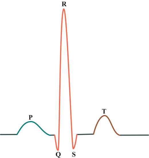
The electrocardiogram provides information about the heart rhythm and heart rate. If the heart beats evenly, a characteristic pattern of spikes and waves results. The sections between the waves and spikes are called the route:
- P wave: The P wave shows how excitation spreads across the heart’s atria as they contract and pump blood into the heart’s chambers (ventricles). Then they relax again.
- QRS complex: The most complex wave in the ECG begins with the Q wave and ends with the S wave. The so-called QRS complex represents the excitation of the heart’s chambers as they contract and pump blood into the body. The QRS complex is particularly important for diagnosing heart problems.
- T wave: The T wave indicates the recovery of the ventricles as they relax and prepare for the next heartbeat.
-
© ScientificStock – stock.adobe.com
The evaluation of an ECG is the responsibility of doctors. A look at the waves and spikes in the ECG curve is usually enough to make a diagnosis. Deviations in the excitation pattern indicate various cardiac disorders. If the Q wave is wider and deeper than usual, this indicates a heart attack. Other important measurements include the ST segment, the PQ interval or the QT duration.
Resting, exercise and long-term ECG: What’s the difference?
The resting ECG is carried out in the doctor’s office and is done while lying down. However, the resting ECG does not provide meaningful information for every heart disease. Some symptoms only become apparent after physical exertion. In this case, a stress ECG may be indicated. This examination of the cardiac current curve is carried out while patients are doing physical activity, for example on the bicycle ergometer.
Even temporary cardiac arrhythmias may escape the resting ECG and may require a long-term ECG. The measurement takes place over a period of around 24 hours. In addition, a long-term ECG is also used to monitor therapy if cardiac arrhythmias are diagnosed or to check after implantation of a pacemaker.
Inflammation of myocardium: These symptoms can be warning signs
