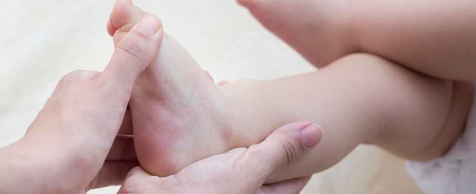
Usually clubfoot is congenital: the newborn's foot is strongly turned inwards. If left untreated, this leads to major problems. However, if the misalignment is corrected in the first few weeks of life with bandages, splints or an operation, those affected can later live normally.
- Every 1000th baby is born with a club foot.
- © iStock.com/Petardj
Article content at a glance:
Recognize skin diseases in children

How does a clubfoot come about?
How a clubfoot (Pes equinovarus) is formed is still discussed among medical professionals. On the one hand, there are genetic defects that lead to a malformation of the skeleton and can also affect the foot. Therefore club feet are more common in some families. On the other hand, doctors suspect muscle problems as the cause – in some babies the muscles of the legs do not grow adequately in the womb.
Causes for this can be:
Too little amniotic fluid: If there is not enough amniotic fluid in the uterus for a long time, the legs do not have enough space to develop straight.
Lack of oxygen: A lack of oxygen can also lead to deformed limbs.
Unfavorable location: If the fetus lies awkwardly in the uterus, the legs may not grow well.
A clubfoot is often made up of several misalignments such as sickle foot, pointed foot and hollow foot. Muscles, bones, and tendons do not grow evenly. This affects the functionality of the foot.
Clubfoot can also develop in healthy children and adults: in the past, it was often triggered by polio. Today, thanks to vaccination, this disease no longer occurs in Europe and America. Injuries, nerve damage, and neurological disorders can also lead to clubfoot. However, acquired malpositions only make up a small part of club feet.
How do you recognize a club foot?
In severe cases, even laypeople immediately see the problem: the newborn's feet point clearly inward, the joints appear to be kinked. Often only one foot is affected, sometimes both. Around one in 1,000 children is born with a club foot. Boys suffer from this deformity about twice as often as girls. By turning the foot, the outer side faces down. In extreme cases, those affected even walk on the back of their feet. The malposition is usually associated with malformations both in the joint (especially in the ankle) and in the bones, tendons and ligaments. The lower leg muscles are also usually deformed in clubfoot. The Achilles tendon is often shortened in clubfoot.
Diagnosis of misalignment of the foot
Usually a clubfoot is noticed during the U1 examination immediately after birth. The doctor checks how far the foot can be moved and rotated in the different directions. He also checks the knee and hip joint as well as blood circulation. He pays attention to the training of the calves and muscles. It is important to distinguish between a baby whose foot has organic malformations and one who turns its foot inward without such malformations.
An x-ray is only necessary if the foot appears severely impaired. Then the doctor can use the X-ray to see whether there is an elevated heel. However, this is usually only the case with older children. You then just walk on tiptoe. Magnetic resonance imaging is required if there is suspicion of fused foot bones (coalition talare). In the case of an adult, dynamic foot pressure measurement can also be useful (pedography), in which it is measured at which point pressure is on the foot.
Ten tips for joint pain

Therapy for a clubfoot
In order to get the foot as well and carefully as possible into the correct position, it must be treated as early as possible. Doctors recommend applying a so-called plaster cast reduction in the first few days of life. The foot is moved in the right direction with the help of a padded plaster of paris. Almost every week, the cast is readjusted to get the foot used to a normal position. Usually the foot has achieved this goal after three to eight different plasters.
Treatment of a clubfoot according to Ponseti
The Ponseti treatment, which is carried out in three steps, has prevailed today:
- Redression (roughly: pushing back)
- Tenotomy (minor surgery)
- Denis Brown splint
First, within the first few weeks of life, the foot is brought into a normal position by the plaster of paris reduction. As soon as the joint has reached an external rotation (mobility) of around 70 degrees, the tenotomy follows: the doctor cuts the Achilles tendon under general or local anesthesia. This usually makes the joint fully mobile again. The foot is then held in the desired position with a plaster cast for about three weeks. The Achilles tendon grows back together completely and properly within a few weeks.
In the third phase of treatment, the child is put on a Denis-Browne splint. Two shoes are attached to a metal rail and can be fixed at the desired angle. The healthy foot is usually determined to be 40 degrees external rotation, the clubfoot to 70 degrees. The baby has to wear these shoes all the time for the first three months. Subsequently, until the age of four or five, he or she has to put on the Denis Brown brace for 14 to 16 hours a day, i.e. at night and during lunch. It is intended to prevent the foot from turning back into its original position (relapse).
Course and prognosis of a clubfoot
If a clubfoot is treated immediately after birth and the parents carry out the treatment consistently, the chances of recovery are very good. However, many small children find the splint annoying as they get older because they are restricted in their movement. Since they do not yet understand the meaning, the parents have to master a great challenge here. Accompanying physiotherapy, which is also carried out regularly by the parents at home, supports the success of the treatment.
Nevertheless, in some cases a relapse (relapse) can occur. If an operation is then necessary, there are general operational risks such as wound healing disorders, wound infections or vascular injuries.
Children whose clubfoot has been successfully treated often later have differently sized feet and accordingly need different shoes. Often they have one calf thinner than the other. The aim of every treatment is to enable those affected to run and exercise normally. In most cases it works.
Preventing a clubfoot
Parents cannot really prevent clubfoot. However, pregnant women should attend all preventive examinations in order to identify any deficits immediately. A healthy lifestyle is also recommended: in particular, a varied diet, exercise in the fresh air and avoiding alcohol and tobacco ensure that the baby can develop well in the stomach. Adults who experience neurological problems, particularly in the legs, should seek good early treatment to avoid clubfoot.